Hartmuth B. Bittner, MD
Theodore N. Pappas, MD
Background
The term gastroesophageal reflux disease (GERD) has been introduced to represent a wide spectrum of damage to the oropharynx, larynx, respiratory tissue, and esophagus clinically recognized by heartburn.
In 1951, Allison (1) coined the term reflux esphagitis. He also described the relationship of hiatal hernia and reflux esophagitis. In 1956, Rudolph Nissen introduced the fundoplication as a simple operative correction of reflux esophagitis (2). The operation was performed over a large esophageal bougie through a left subcostal incision or a lower left thoracotomy (figures 1 through 3). Nissen's technique was to mobilize the abdominal segment of the esophagus and the lesser curvature of the stomach by division of the gastrohepatic ligament. Almost at the same time as Nissen's introduction of an antireflux procedure, the Belsey Mark IV and Hill gastropexy were described (figures 4 through 8). The Belsey Mark IV repair is performed through a left sixth interspace thoracotomy to create a 240° fundic wrap (3). The technical objectives of the Hill procedure (4) involve utilization of the anterior and posterior phrenoesophageal bundles to anchor the gastroesophageal junction posteriorly to the preaortic fascia and the median arcuate ligament (posterior gastropexy). In 1963, Toupe (5) modified the Nissen fundoplication such that he created a posterior 270° fundic wrap (figure 9).
In 1991, Geagea (6) carried out the first Nissen fundoplication using a solely laparoscopic technique in a 43-year-old woman with severe reflux esophagitis. The important part of the operation was the creation of the 360° fundus wrap by laparoscopic technique. Seromuscular gastric silk sutures were used to secure the wrap. The vagi nerves were preserved and ordinarily included with the fundic wrap.
Bittner and coworkers (7) reported a patient series who underwent the Nissen fundoplication with the innovative technique and under laparoscopic guidance for GERD. This technique essentially mimicked the traditional open Nissen procedure, and a success rate of greater than 90% was achieved.
Nissen fundoplication under laparoscopic guidance offers an alternative form of surgical therapy for patients with GERD. The laparoscopic technique avoids many of the disadvantages of the traditional approach such as a large abdominal incision, associated postoperative discomfort, and prolonged recovery periods.
Anatomy
The esophagus comprises the cervical, thoracic and abdominal segments. The point of origin corresponds to the caudal border of the cricoid cartilage and the lower margin of the cricopharyngeus muscle at the level of the sixth cervical vertebral body. The esophagus extends downward through the superior and posterior mediastina of the thorax. It passes through the esophageal hiatus of the diaphragm to join the cardia of the stomach about the level of the tenth thoracic vertebra.
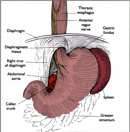
Fig. 10
The accompanying structures on its course through the hiatus are the anterior and posterior vagal trunks (figure 10) In its short abdominal portion (25 to 30 cm in length), the esophagus lies upon the diaphragm with the esophageal impression of the left lobe of the liver applied to its anterior aspect. The length of the intra-abdominal segment in the normal state averages 1.5 to 2 cm.
The lower esophagus segment is subdivided into the supradiaphragmatic portion, inferior esophageal constriction, vestibule, and cardia. This segment is supplied by an ascending branch of the left gastric artery. Venous drainage of that segment enters the coronary vein and pericardial venous plexus below the diaphragm. Preganglionic fibers of the vagus nerve synapse with ganglion cells in the myenteric plexus of the esophagus. Sympathetic innervation occurs from the thoracic ganglia.
The anatomy and physiology of the gastroesophageal junction and of the esophageal hiatus is essential to the understanding of reflux mechanisms and antireflux procedures. The muscle fibers surrounding the esophagus and creating the hiatus are mainly from the right crus of the diaphragm (figure 10). In addition, several layers that separate the thoracic and the abdominal cavity support the esophageal hiatus.
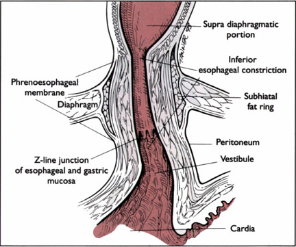
Fig. 11
The most important structure is the phrenoesophageal membrane, a condensation of endoabdominal and endothoracic fascia (figure 11). This membrane anchors the esophagus within the hiatus. In most individuals this membrane inserts 3.3 cm above the junction of the tubularesophagus with the stomach. Part of the problem of defining the gastroesophageal junction is the fact that the internal junction does not coincide with the external junction. Gahagan (8) describes this region as follows: "the external junction the termination of the tube, the esophagus and the beginning of a pouch, the stomach lies 1 cm below the internal junction, the mucosal boundary."
Pathophysiology, Presentation, and Differential Diagnosis
It is now recognized that although there is no anatomical lower esophageal sphincter in humans, a physiologic sphincter mechanism exists and extends over the terminal 1 to 4 cm of the esophagus. Manometrically, this specialized segment represents a zone of high pressure referred to as the lower esophageal sphincter (LES). In the resting state, the LES is contracted and its mean pressure is 13 mm of mercury. The overall length of the LES is 3.6 cm on average, 2 cm as the intra-abdominal portion and the remainder in the thoracic cavity. The control of gastroesophageal reflux is achieved by the following mechanisms: intrinsic muscle tone of the LES, intra-abdominal esophageal segment, abrupt opening of the narrowing swallowing tube into dilated gastric pouch (la Place law), and normal gastric emptying. The functional state of the LES determines the development of GERD. Patients with an LES pressure of less than 5 mm of mercury or an abdominal portion smaller than 1 cm present with GERD in 90% of cases (9).
Hiebert and Belsey's (10) observations clearly showed that pathologic reflux and hiatal hernia were separate entities. Approximately 80% of patients with GERD have a radiographically demonstrable axial hernia. When radiographic maneuvers are applied to diagnose a hiatal hernia, approximately only 5% of such individuals have pathologic gastroesophageal reflux. Presumably, this is because the phrenoesophagela membrane insertion still leaves an adequate segment of distal esophagus exposed to abdominal pressure. Reflux is prevented when there is a 10-cm water column abdominothoracic pressure difference. (11)
Although delayed gastric emptying has a strong correlation with severe reflux and esophagitis, it is not known whether the gastric emptying delay proceeds the pathologic reflux or whether esophageal inflammation from reflux esophagitis causes vagal nerve dysfunction and inhibits gastric emptying.
Twenty-four-hour intraesophageal pH monitoring documents that normal individuals have physiological reflux after meals. Seven percent experience heartburn every day and 36% of healthy individuals experience heartburn once a month. Pathologic degrees of reflux are diagnosed when reflux becomes prolonged or occurs throughout the day or at night. Esophagitis as a complication of reflux develops when gastric acid or pancreaticobiliary secretions reach the esophagus with increased frequency and a failure of protective esophageal mechanisms occurs. An important aspect in the protection against noxious effects of gastroesophageal reflux is coordinated peristaltic clearing and secondary peristalsis of the esophagus that is triggered by distention or irritation of the mucosa of the distal esophagus. When abnormal reflux occurs, the damage to the esophageal mucosa may range from none to the development of a severe peptic stricture (figure 12). The degree of damage to the esophagus is graded as I (erythema of the mucosa without ulcerations), II (ulcerations), III (chronic ulcerations with fibrosis), and IV (stricture) (13). Table 1 summarized the symptoms and complications in patients who present with symptomatic GERD. The most common differential diagnoses, such as cholelithiasis, peptic ulcer disease, gastritis, esophageal motor disease, and angina pectoris are demonstrated in (figure 13).
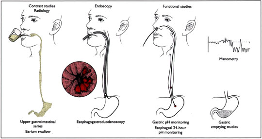
Figure 14: Diagnostic studies in patients with gastroesophageal reflux disease
Diagnostic studies to define GERD and to evaluate patients for laparoscopic Nissen fundoplication include manometery of the esophagus, 24-hours pH monitoring, and esophagogastroduodenoscopy with biopsies (figure 14). A mechanical sphincter defect is defined by a resting pressure less than 6 mm Hg or less than 2 cm in length (14).The indications for laparoscopic Nissen fundoplication are identical to the open antireflux procedures. Primary indications for surgery are medical management failure, need for long-term omeprazole treatment, or complications of GERD like high-grade esophagitis, stricture or Barrett's esophagus (figure 12 and figure 15).
Surgical Technique
The Nissen fundoplication, which is done laparoscopically, is identical to the one via the open technique (figures 16 to 21). For the laparoscopic construction of the fundoplication, pledgets can be used between the esophagus and stomach as described by DeMeester and coworkers (5) in 1986. A Òfloppy NissenÓ wrap of approximately 2 to 3 cm is created over a 58- to 60- French bougie to parallel the open technique as close as possible. (figures 22 to 28; figures 29 and 30 illustrate an alternative procedure). The short gastric vessels are not ligated or divided for the mobilization of the gastric fundus. Grural repair or approximation is not undertaken. Anterior and posterior vagus nerves are included in the wrap. The Hasson cannula is used in all patients with a history of previous abdominal surgery.
Set-Up
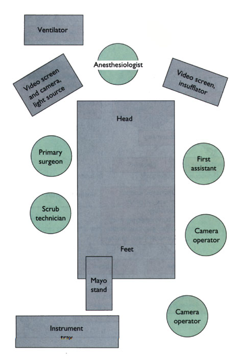
Fig 16
Operative set-up for laparoscopic Nissen fundopliction. Foley and nasogastric catheters are placed after induction of general anesthesia. the patient is then placed in the 30° Trendelenburg position with the arms tucked at the sides. A pneumoperitoneum is established with a Veress needle inserted into the peritoneal cavity through a 1-cm infraumbilical incision. In patients with previous abdominal surgery the open technique is used and an approximately 2.5-cm incision is made for the introduction of the Hasson cannula. A 10-mm sheath is used for the insertion of the video camera.
Trocar Placement
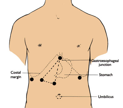
Fig 17
Trocar placement for laparoscopic Nissen fundoplication. The pneumoperitoneum is maintained at 15 mm Hg. In patients with a large abdomen or a long distance between umbilicus and xiphoid process, a supraumbilical incision is made for the insertion of the video camera. Five more trocars are introduced under visual control. A 10-mm trocar in the left upper quadrant is necessary for placement of an endoscopic Babcock clamp. A 10-mm trocar is placed at the upper midline for a fan retractor. A trocar is placed in the far right lateral aspect of the costal margin for placement of a second endoscopic Babcock clamp. The triangle describes the trocar placement for the surgeon operating two handed at the base of the triangle. The trocars should be placed as high as possible to allow them to reach the hiatus. The apex of the triangle faces the gastroesophageal junction.
The Procedure
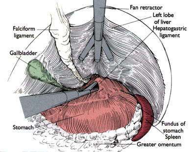
Fig 18
This illlustration demonstrates the view as seen from the 10 mm torcar at the umbilicus. The fan retractor elevates the left lobe of the liver. The hepatogastric ligament is sharply dissected for the mobilization of the gastroesophageal junction.
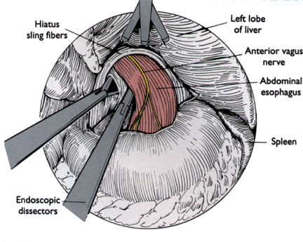
Fig 19
The hepatogastric ligament is opened. The right crus of the diaphragm is then mobilized from the gastroesophageal junction. Care is taken toaccurately distinguish the crus from the esophagus. Moving the nasogastric tube in the esophagus while the gastroesophageal junction is viewed helps to define the anatomy.
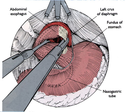
Fig 20
With the esophagus retracted to the patient's right, the left diaphragmaticcrus is dissected, and the space between this side of the esophagus and crura is opened bluntly. With the esophagus retracted upward, the retroesophageal space is dissected under direct vision. Care is taken not to enter the pleural space.
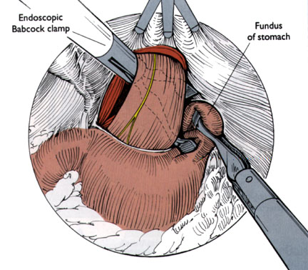
Fig 21
An endoscopic Babcock clamp if placed from the most lateral right trocar and is passed behind the gastroesophageal junction. The placement of instruments
in this area is done under direct vision. The fundus of the stomach is grasped with a second Babcock clamp from the left side and passed behind the gastroesophageal junction. The nasogastric tube is replaced with a 58 or 60 French bougie.
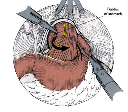
Fig 22
The right endoscopic Babcock clamp is gently pulling the fundus of the stomach across the back of the gastroesophageal junction. With the same segment of stomach that was being passed around the back of the esophagus, the 360° wrap is created.
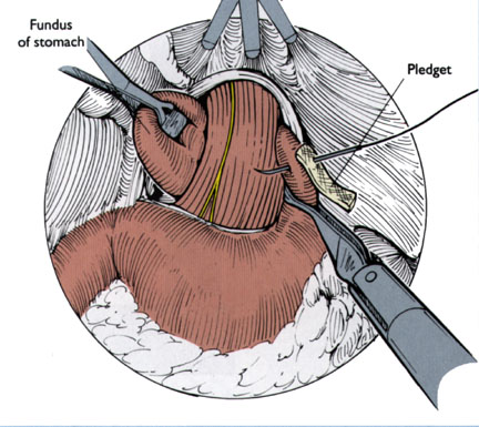
Fig 23
Pledgets are then passed into the abdomen. Pledgets are used at each interface of stomach and esophagus to support a zero nonabsorbable suture. The anterior and posterior vagus nerves are included in the wrap.
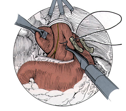
Fig 24
Four pledgets are used. At first the superior suture is placed, usually from the left side to the right side. Seromuscular stitches are performed.
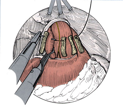
Fig 25
The suture is passed from the left side through pledget, stomach, pledget, esophagus, pledget, stomach and pledget.
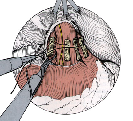
Fig. 26
The suture is then brought out through the same 10 mm trocar through which it was introduced. An extracorporal knot is tied, which is introduced into the peritoneal cavity and slid down with a pusher bar.
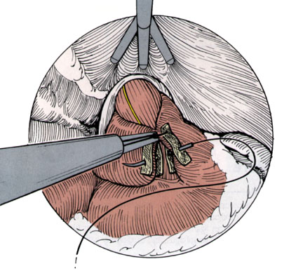
Fig. 27
A second suture is placed inferior to the previous one, approximately 1 to 1.5 cm apart, using an identical technique.
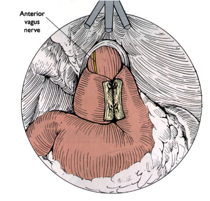
Fig. 28
The complete 360° wrap is demonstrated. The length of the wrap is approximately 2 cm and as demonstrated left, created over a large bougie to create a loose "floppy Nissen". Trocars are then removed, the pneumoperitoneum released, the incisions closed with interrupted number 1 polyglactin sutures, and incision dressings are applied.
Alternative Set-up
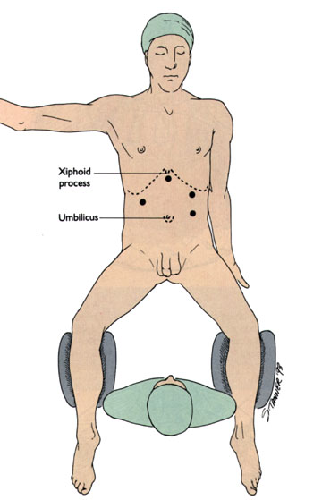
Fig 29
The surgeon is positioned between the legs of the patient. The patient is in lithotomy and reverse (20° to 30°) Trendelenburg position. Five trocars are placed high in the epigastrium. The video camera is introduced through the left upper quadrant trocar.
Alternative Procedure
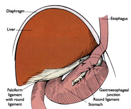
Fig 30
Alternative laproscopic antireflux procedure, laparoscopic ligamentum teres (round ligament) cardiopexy. The procedure consists of three sequential parts: mobilization of the teres ligament with preservation of its blood supply, mobilization of the abdominal esophagus, and cardiopexy with the teres ligament sling [17].
Results
Dallemagne and coworkers [16] reported on the first series of 12 patients who underwent laparoscopic Nissen fundoplication. Three of the 12 patients required conversion to open laparotomy for equipment failure or bleeding encountered during dissection of the short gastric vessels. The remaining nine patients reported complete relief of symptoms. Laparoscopic reduction, crural repair, and fundoplication of a large hiatal hernia was performed by Cushieri and coworkers [18] in eight patients. Crural repair and fundoplication was done by continuous suture technique. The average operating time was 3 hours. The postoperative complications were cervical emphysema (three patients), left pneumothorax (one patient), and transient dysphagia (one patient). All had no symptoms and absence of acid reflux 3 months postoperatively.
Bittner and coworkers [7] in 1993 laparoscopically explored 35 patients for Nissen fundoplicaton. There was a 0% mortality rate and the total hospital morbidity rate was 25.7% (nine of 35 patients). Five patients required conversion to open Nissen fundoplication, three due to hemodynamic instability secondary to presumed pneumothorax, a transverse colotomy in one patient, and a distal esophageal perforation in another patient. The total surgical time o laparoscopic Nissen fundoplication was 109 minutes on average. The majority of the patients were discharged on postoperative day 2 or 3. Three patients developed severe dysphagia due to a tight wrap, whereby two patients required dilation for resolution of their symptoms, and the other patient's symptoms resolved spontaneously. Overall 26 of the 30 patients who underwent the laparoscopic Nissen fundoplication (87%) described the outcome of the operation as "excellent" and "good". All patients returned to baseline activities and full employment within 14 days after the operation. The clinical assessment and satisfaction scores of the 30 patients who successfully underwent laparoscopic Nissen fundoplication are shown in table 2.
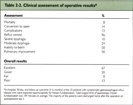
In conclusion, laparoscopic Nissen fundoplication can be performed safely and is an effective antireflux procedure. It has the same good results as standard Nissen fundoplication. The best results are obtained in carefully selected patients who present predominantly with regurgitation and heartburn. As in many other laparoscopic procedures, a shorter hospital stay and an earlier return to baseline activities is recognized. Total costs are less compared with the standard technique due to a decreased hospital stay and earlier return to work [19}.
References
Used by permission of Current Medicine, Philadelphia.
Copyright © 1996 by Current Medicine. All Rights Reserved.
Atlas of Laparoscopic Surgery is distributed by:
Church Livingston
Attn: Department - JW
650 Avenue of the Americas
New York, NY 10114-0638
Tel: 800-553-5426
Fax: 708-983-5576
Contact webteam