IMAGE-GUIDED BREAST BIOPSY: THE ABBI* SYSTEM
by William E. Kelley, M.D.
 Stereotactic core needle biopsy is one procedure for
tissue diagnosis of a nonpalpable breast lesion presenting as a suspicious
finding on mammography. Ultrasound-guided needle biopsy is a cost effective
alternative, but is more operator-dependent, with a reported accuracy of
47-91%. Stereotactic core biopsy is minimally invasive, requiring only a
4mm incision, and is safe. It has a reported accuracy of 98% in experienced
hands, which equals the results of surgical series.
 Stereotactic Technology
United States Surgical Corporation has developed a new technology which
permits sampling of a mammographic lesion while preserving the histologic
architecture for complete, traditional pathologic analysis. The Advanced
Breast Biopsy Instrumentation, or ABBI* System utilizes the same stereotactic
guidance system as core needle biopsy. The ABBI* system was developed in conjunction with
Lorad, a Division of ThermoTrex Medical Corporation.
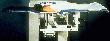 ABBI* Table
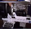 Compress Breast
Stereotactic x-rays are taken allowing the physician to visualize and target the non-palpable lesion's location to within 1mm of accuracy.
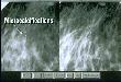 Stereotactic X-rays
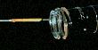
A localizing needle is guided to the precise location of the lesion by digital stereotactic X-rays through an incision.
 Localizing Needle
The position of the needle is verified by digital stereotactic X-rays, and a T-fastener is then deployed, thus fixing the lesion in place. The position of the T-fastener is then confirmed with another stereotactic X-ray, completing the localization sequence.
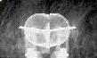 T-fastener
The biopsy sequence begins with further infiltration of local anesthesia, and the incision is extended to a length 3 mm longer than the diameter of the biopsy cannula. If the lesion is deep in the breast, the physician undermines the superficial tissues with scalpel dissection to reduce the size of the ultimate specimen. A cylindrical cannula is then advanced over the needle and T-fastener while a motorized, oscillating, circular cutting blade incises the breast tissue.
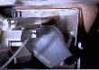 Biopsy Sequence
The cannula is advanced beyond the lesion to a predetermined depth, and another stereotactic X-ray is taken to confirm that a sample has been captured within the cannula. At every step, the precise location of the device and the lesion are verified by digital X-rays.
 Cannula Sample
A cautery snare then cuts across the deep end of the cannula, completing the biopsy, and the cannula and specimen are removed.
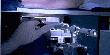 Cautery Snare
When the specimen has been removed, a completion mammogram is taken before the patient is taken out of compression.
 Digital X-rays
A specimen X-ray is performed to confirm that the targeted tissue is contained within the sample.
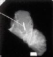 Specimen X-ray
The patient is then rolled supine and the biopsy site is inspected. The deep portion of the biopsy is marked with a clip under direct vision. Bleeding is an infrequent problem and is easily controlled with cautery or clips or sutures.
There are four cannula sizes available: 5, 10, 15 and 20 mm.


The smallest ABBI* unit removes a specimen smaller than a pencil eraser and the largest unit removes a specimen the size of half a wine bottle cork. The pathologist makes cross sections, converting the specimen into a "stack of coins." The architecture of the lesion is seen in cross section. The physician chooses the cannula size consistent with the size of the sample desired.
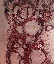 Pathology Specimen
ABBI* procedure: no second diagnostic procedures have been needed.
The physician and patient have multiple options for tissue diagnosis of a mammogram finding. Image-guided, minimally invasive biopsies have both cosmetic and economic advantages. The size of the incision is predictable for each cannula and heals remarkably well, probably due to the atraumatic nature of the procedure, lack of tissue damage from retractors, and absence of talc contamination. Core biopsy incisions are smaller, measuring 4 mm as compared with 8, 13, 18, or 23 mm for the ABBI* biopsies.
Most ABBI* patients have the security of knowing that the targeted tissue is no longer seen on X-ray, which is a very important issue for some women. For other women, the 4 mm incision is a more important factor and they are willing to accept an 18% rebiopsy rate and the need to follow their residual mammographic lesion closely. The patient who receives a malignant diagnosis following ABBI* biopsy can usually be informed of her prognosis and can hear a full and weighted discussion of her therapeutic options, since the physician has the advantage of full and detailed pathologic examination. After a lengthy discussion of the advantages and disadvantages of the image-guided biopsy options, the patient can make an informed decision and choose the best option for her.
On a final note, it must be emphasized that the ABBI* biopsy is a diagnostic procedure. Randomized prospective studies will be needed with long term follow-up results before the ABBI* procedure can be performed as a therapeutic procedure.
BIBLIOGRAPHY
- Berg WA Ruban RH, Kumar D, el al. Lessons from mammographic-histopathologic correlation of large-core needle breast biopsy. Radio Graphics, 1996; 16: 1111-1130.
- Dershaw DD, Morris EA, Liberman L, et al. Nondiagnostic stereotactic core breast biopsy: Results of rebiopsy. Radiology, 1996; 198: 323-325.
- Lee CH, Egglin TK, Philpotts L, et al. Cost-effective of stereotactic core needle biopsy: Analysis by means of mammographic findings. Radiology, 1997: 202: 849-854.

