Dr. Saye was the assisting surgeon on the first endoscopic cholecystecomy
procedure performed in the U.S., and has gone on to complete almost 500
LDH procedures. He recently led the development of a new minimally invasive
therapy, laparoscopic Doderlein hysterectomy, and has improved on numerous
other laparoscopic gynecologic procedures. He has been in private practice
in Marietta since 1971.
LAPAROCOPIC DODERLEIN HYSTERECTOMY WITH BILATERAL SALPINGO-OOPHORECTOMY William B. Saye, MD, FACOG, FACS
The Procedure: Laparoscopic Portion
The laparoscopic Doderlein hysterectomy (LDH) is a viable alternative
to conventional abdominal hysterectomy. Over the past 4 years, I found that
96% of the abdominal hysterectomies performed at our facility could be converted
to the LDH. All hysterectomies that could be performed vaginally, and all
hysterectomies that were found not to match the profile established for
safe vaginal hysterectomies were included in the LDH group.
Patients who conform to the standard preoperative protocol (after obtaining
a complete history, a physical examination, and a recent Pap smear) are
selected as candidates for the LDH. These patients are informed that the
operation could be converted to a conventional open procedure if the dissection
should prove to be too difficult or if an emergency arises necessitating
the conversion. The surgeon's inability to totally predetermine anatomical
anomalies is also a bona fide reason for converting the LDH to an open procedure.
The procedure is begun by performing a pelvic examination after placing
the patient in the dorsal lithotomy posistion. A tenaculum is placed on
the anterior cervical lip, and a uterine manipulator inserted into the endo-cervical
canal. An elliptical periumbilical incision is made. The subcutaneous tissue,
fascia and peritoneum are entered under direct vision, and a 12 mm VERSAPORT*
trocar is placed in the umbilical site.
Plate 1
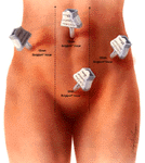
The peritoneal cavity is insufflated to an intraperitoneal pressure
of 15 mm Hg. A 5 mm trocar is then placed in the lower midline, 2 fingerbreadths
above the symphysis pubis, under direct vision. A 12 mm trocar is then placed
in the left lower quadrant at or below the level of the umbilicus, depending
upon the size of the uterus. If the uterus is large, this trocar is placed
at the level of the umbilicus. If the uterus is smaller than the size of
a 12-week uterine pregnancy, the trocar is placed approximately 1 inch below
the umbilicus. This trocar is placed lateral to the rectus muscle, care
being taken not to injure the inferior epigastric vessels as the trocar
is inserted.
The camera is then moved to the left lower quadrant port, and attention
is turned to the right lower quadrant or the level of the umbilicus where
a third 12 mm VERSAPORT* trocar is placed at the fourth trocar site, lateral
to the rectus muscle and lateral to the inferior epigastric vessels. Care
again must be taken not to injure any vascular structures of injure the
bowel upon insertion of the trocar. SURGIGRIP* sleeves are used on all the
disposable trocars, and VERSAPORT* disposable trocars with safety shields
are used in all ports.
Prior to insertion of the 12mm trocars, a 2-0 POLYSORB* suture is placed
in the skin incision by means of an ENDO CLOSE* needle to close the fascia
and peritoneum at the termination of the procedure.
The patient is placed in an exaggerated Trendelenburg position; shoulder
braces are not used however, because of the risk of injury to neurovascular
structures in the upper extremities. Three ENDO GRASP* instruments are then
inserted through the trocar sites with the camera in the umbilical port,
and in a systematic fashion, beginning at the ileocecal valve, the small
bowel is examined, removed from the pelvic cavity and placed in the right
upper quadrant. The appendix is also visualized, as are the liver, gallbladder
and all of the upper abdominal structures to rule out any pathology.
After the bowel has been satisfactorily removed from the pelvis, both
ureters are identified prior to beginning dissection. Any ancillary dissection
required- including adhesions of bowel to pelvic structures or of ovaries
and tubes to the uterus or to the lateral pelvic wall - are dissected free
at this time. Any endometrial implants are also treated with the fiberTome
laser prior to the initiation of the operative procedure per se.
Plate 2
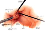
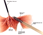
If the ovaries are to be removed, after the ureter has been identified,
a MULTIFIRE ENDO GIA* stapler with a gray cartridge is placed through the
right lower quadrant port to transect the right infundibulopelvic ligament.
The ureter is again identified after the MULTIFIRE ENDO GIA* 30 cartridge has been
applied to the infundibulopelvic ligament.
Plate 3
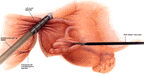
After the infundibulopelvic ligament has been transected, a second MULTIFIRE
ENDOGIA* 30 cartridge is used to transect the round ligament. This is usually
a white ENDO GIA* 30 cartridge, and it is also placed through the ipsilateral
port to position the dissection close to the uterus and away from the ureter
on the side of dissection. Again, the ureter must be examined before the
stapler is applied to the paraovarian tissue and round ligament, and after
the ENDO GIA* 30 cartridge has been closed. A third ENDO GIA* 30 cartridge
is usually required to transect the broad ligament down to approximately
5 mm above the uterine artery. This is also a gray cartridge. This may sometimes
be replaced in the umbilical port of the camera is moved to the right or
the left lower quadrant before the application of the cartridge, parallel
to the uterus, and as close to the uterus as possible.
Plate 4
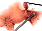
Plate 5
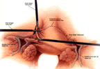
The same procedure is carried out to transect the left infundibulopelvic
down to and including the round ligament and the upper portion of the broad
ligament.
If the ovaries are to be spared, the camera is placed in the right lower
quadrant or the right upper quadrant, depending on where the trocar is placed.
The uteroovarian pedicles are transected on both sides with a white MULTIFIRE
ENDOGIA* 30 cartridge. A second application of the MULTIFIRE ENDO GIA* 30
cartridge is then used to transect the upper portion of the broad ligament.
This is usually a gray cartridge.
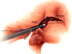
At this point in the operation, a bladder flap is developed in the lower
uterine segment with the use of sharp dissection, using the ENDO SHEARS*
instrument to develop the bladder flap, (we have found that aqua dissection
is very seldom required to create the bladder flap. With careful sharp dissection
and pneumodissection of the peritoneum with CO2 used to insufflate the peritoneum,
the bladder flap is created with very little effort.)
The bladder is dissected off the lower uterine segment and the cervix
until the endopelvic fascia overlying the cervix is identified. This frequently
requires the use of an ENDO PEANUT* in order to bluntly dissect the bladder
from the cervix. After the bladder has been dissected from the lower uterine
segment, careful inspection is carried out on each side to visualize the
ureter. The last application of the MULTIFIRE ENDO GIA* 30 stapler is on
each side of the uterus parallel to the cervix, with the dissection stopping
approximately 5 mm above the uterine artery on each side. We believe that
the uterine artery should not be routinely transected during the laparoscopic
portion of the operation. We find that it is much safer to transect the
uterine artery after the uterus has been delivered through the anterior
cul-de-sac while the vaginal dissection is being performed. Careful measurement
of the MULTIFIRE ENDO GIA* 30 cartridge reveals that it spans 9 mm from the
inside of the cartridge to the outside row of staples. Quite often the operator
will not have 9 mm of space between the cervix and the ureter, and in our
opinion, application of the MULTIFIRE ENDO GIA* 30 stapler below the uterine
artery is sometimes risky. If the decision is made to staple the uterine
arteries laparoscopically, a MULTIFIRE ENDO TA* 30 stapler is used and the
incisions are made on the specimen side of the 3 rows of staples after the
uterus is delivered into the inferior cul-de-sac.
At this juncture, attention is turned to the vaginal portion of the
procedure, which is performed with the Doderle in technique.
The Procedure: Doderlein Portion
After the laparoscopic portion of the Doderlein hysterectomy is completed
with the use of the MULTIFIRE ENDO GIA *30 stapler, we discontinue the dissection
approximately 5 mm above the uterine artery on both sides. We then turn
our attention to the vaginal portion of the procedure.
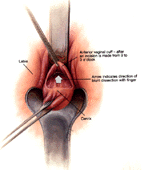
An incision is made on the anterior cervical lip from 9 to 3 o'clock. With
the use of sharp dissection, the dissection plane is effected between the
cervix and the bladder. With the use of blunt dissection, the bladder is
separated from the cervix. Bear in mind that the peritoneum has already
been incised during the laparoscopic portion of the operation, so the peritoneum
does not have to be identified at this time. The operator will feel his/her
index finger pop into the peritoneal cavity as it is entered. This can be
visualized on the monitor if the laparoscopic camera has been kept in place.
At this point, a right-angle Heaney retractor is inserted into the anterior
cul-de-sac. Prior to completion of the laparoscopic portion of the operation,
it is very helpful if the uterus is scored in the midline with a suture
placed during the laparoscopic portion of dissection. This will enable the
operator to identify the midline of the uterus in the anterior surface all
the way up to the fundus of the uterus.
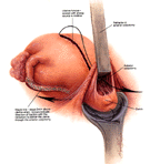
After entering the cul-de-sac with the use of a Lahey thyroid tenaculum,
the operator moves up the anterior portion of the uterus in the midline
all the way to the fundus, which has been marked during the laparoscopic
dissection. As traction is placed on the anterior surface of the uterus
with the left hand, countertraction is placed on the posterior cervix with
the right hand. Pressure is directed toward the rectum with the right hand,
and the fundus of the uterus is pulled into the anterior colpotomy with
the left hand. This creates a see-saw effect, thereby delivering the fundus
through the anterior cul-de-sac.
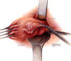
Once the fundus has been delivered through the anterior cul-de-sac,
the tenaculum is released form the posterior cervix and a lateral retractor
is placed into the lateral vaginal fornix to the operator's right side (the
patient's left side). With a Heaney clamp, the staple line is continued
toward the cervix, thereby grasping the uterine artery and the upper portion
of the cardinal ligament. This tissue is then divided sharply with scissors
and a suture ligature is placed in this pedicle. A thyroid tenaculum is
again placed in the posterior cervix, and using a Heaney clamp, the lower
portions of the cardinal ligament and the uterosacral ligament are grasped
between the jaws of the Heaney clamp. This provides vaginal mucosa against
the lower jaw of the Heaney forceps and peritoneal surface on the upper
jaw of the Heaney clamp. This tissue is then transected with scissors. A
Heaney suture using size 2-0 POLYSORB* suture is placed in this pedicle,
and the pedicle is held with a hemostat for identification of the uterosacral
ligament. The tenaculum is then removed from the cervix.
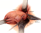
A lateral retractor is placed in the patient's right lateral vaginal
vault. A Heaney clamp is then placed across the right uterine artery and
the upper portion of the cardinal ligament. This is transected with scissors
and a suture ligature placed in this area using size 2-0 POLYSORB* suture.
The thyroid tenaculum is then reapplied to the posterior lip of the cervix,
and a Heaney clamp is placed on the uterosacral ligament and the lower portion
of the cardinal ligament, thereby incorporating the uterosacral ligament
into the vaginal cuff as it was previously performed on the contralateral
side. This pedicle is suture ligated with size 2-0 POLYSORB* suture.
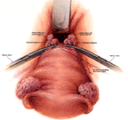
Two Heaney clamps are then placed across the remaining portion of the vaginal
mucosa, and the cervix is amputated from the vagina. A running, interlocking
hemostatic suture using size 3-0 POLYSORB* suture is started in the right
angle of the vaginal cuff and continued across the entire posterior vaginal
cuff until the left uterosacral ligament has been incorporated in this suture.
The vaginal cuff is then closed with interrupted size 2-0 POLYSORB* sutures.
The incorporation of the uterosacral ligament in the vaginal mucosa aids
in the support of the vaginal vault and decreases the chance of vaginal
vault prolapse following the operation.
Notice should be taken that only the anterior colpotomy was performed
with the delivery of the uterine fundus through the anterior colpotomy.
This negates the necessity for a posterior colpotomy in order to complete
the operation. Thus the integrity of the posterior cul-de-sac is maintained,
thereby decreasing the possibility of an enterocele following the Doderlein
technique of laparoscopically assisted vaginal hysterectomy.
The suture material used throughout the completion of the vaginal portion
of this procedure is size 2-0 POLYSORB* synthetic absorbable suture which
is manufactured and sold by United States Surgical Corporation.
Following this, a Foley catheter is placed in the bladder and attention
is again turned to the laparoscopic portion of the procedure where the peritoneal
cavity is re-insufflated and hemostasis is assured. During our first 100
cases, we performed cyctoscopy after completion of the vaginal portion of
the procedure- and after establishing the pneumoperitoneum - to ensure that
the bladder was intact and that efflux from both ureteral orifices was visualized
under direct vision.
Following a successful cyctoscopy, the intraperitoneal pressure is decreased
to 9mm Hg and the ancillary trocar sheaths are removed under direct vision
to ensure hemostasis. Following removal of the ancillary trocar sheaths,
and tying the sutures placed in the fascia of the right and left lower quadrant
trocar sites, the umbilical trocar sheath is then removed after the CO2
and irrigating fluids have been withdrawn from the peritoneal cavity. The
fascia of all VERSAPORT* trocar sites is closed with size 2-0 POLYSORB* suture,
and the skin incisions are closed with size 4-0 plain SURGIGUT* subcuticular
sutures.
I hope that this discussion of the Doderlein procedure simplifies the
technique and enables you to complete the operation in a satisfactory manner.
 Link ALTC page
